Correlative MRI/NMR and X-ray measurement (CORREL)
Note: The choice between CORREL and NMR depends on the specific scientific question and the requirements of the intended work. For large, heterogeneous samples and multi-modal analysis, CORREL is recommended. For high-resolution imaging and detailed molecular analysis, NMR is the better choice. Read more ...
Content
sprungmarken_marker_1214
Technology Description (CORREL)
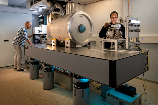
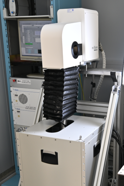
At CORREL, we integrate X-ray imaging (XRI), magnetic resonance imaging (MRI), and nuclear magnetic resonance (NMR) spectroscopy into a single platform, offering a unique correlative approach that combines multi-modal imaging and spectroscopy for sample analysis. CORREL facilitates the simultaneous acquisition of spatio-temporally resolved chemical information (NMR spectroscopy) and structural information (XRI, phase-sensitive XRI, MRI) from a single sample, across various length and time scales specific to each method.
We recommend a preliminary consultation with our team on the following topics before initiating a project:
- Sample concentration and/or quantity;
- Sample size and additional experimental conditions (e.g., temperature, current/voltage, fluids in/out);
- Sample phase;
- Target nuclei to be analyzed;
- Type of samples (e.g., low-absorbing, composite with similar characteristics, degradable, X-ray radiation-sensitive);
- Process to be studied and temporal resolution;
- Desired imaging resolution and contrast requirements.
| Name | Phone | |
|---|---|---|
| Dr. Kunka, Danays | danays kunka ∂does-not-exist.kit edu | |
| Dr. Last, Arndt | arndt last ∂does-not-exist.kit edu | |
| 1 additional person visible within KIT only. | ||
Details (CORREL)
Features / Characteristics
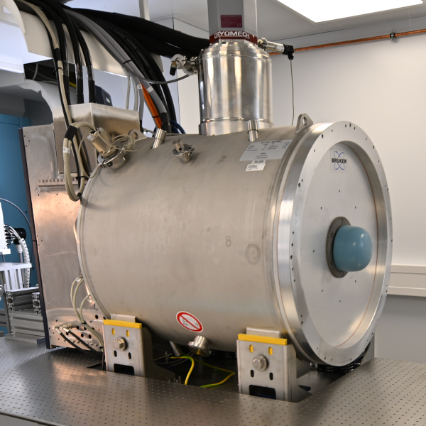
Bruker BioSpec 70/11 System
- Field strength: 7.0 T
- Bore diameter: 105 mm
- Cylindrical homogeneity volume: Diameter x Length 30 x 60 mm
- Homogeneity: peak-peak 4 ppm
- Gradient strength: 740 mT/m
RF Channels
- 1H/19F
- 12C, 7Li, 23Na (other nuclei are possible upon consultation)
X-ray
- Acceleration voltage up to 70 keV
- 250 W max. electron beam power
- X-ray spectrum with Ga Ka-Line at 9.24 keV and In Ka-Line at 24.2 keV
- Source spot size: 20 µm vertical x 80 µm horizontal at 250 W; 10 µm x 10 µm at 10 W
- Resolution in Computed Tomography (CT) imaging: 30 µm voxel size for centimetre size samples, 5 µm voxel size for below 2 mm sample size
Typical measurement methods
- MR imaging and NMR spectroscopy
- Computed Tomography (CT) in about 3 h total exposure time
- Radiography
- Absorption / phase / darkfield contrast imaging using Talbot-Lau gratings
- Hartmann-based multi-contrast spatial harmonic imaging with 250 ms time resolution
Limitations/constraints
- Maximum sample size ~30 mm Ø
- Only non-ferro-magnetic materials in the magnet (if only XRI is needed, magnetic or large samples are possible)
Recommendation: CORREL vs. NMR
Overview of Technologies
CORREL integrates X-ray imaging (XRI), magnetic resonance imaging (MRI), and nuclear magnetic resonance (NMR) spectroscopy into a single platform. This unique correlative approach allows for the simultaneous acquisition of spatio-temporally resolved chemical information (NMR spectroscopy) and structural information (XRI, phase-sensitive XRI, MRI) from a single sample. This makes CORREL particularly useful for multi-modal imaging and spectroscopy across various length and time scales.
NMR focuses on manipulating nuclear spin to reveal molecular structure and dynamics with atomic resolution. It can also generate 3D images of spin density with voxel dimensions down to ~10 µm. NMR can probe a wide variety of materials in different phases (gas, liquid, solution, solid) and operates at high magnetic fields (up to 11.7 T), providing detailed molecular information.
Use Cases
CORREL is best suited for:
- Imaging large samples without the need for cutting
- Localized spectroscopy of large, heterogeneous samples
- Situations where simultaneous or correlated MR + X-ray measurements are beneficial
NMR is ideal for:
- High-resolution imaging and spectroscopy of smaller samples
- Detailed molecular structure and dynamics analysis
- Applications requiring superior spatial resolution and sensitivity
Considerations
- Sample Size: If the sample is large and cannot be cut, CORREL is the preferred choice due to its ability to accommodate larger samples.
- Imaging Quality: For superior imaging quality and spatial resolution, NMR is the better option due to its stronger magnetic field and gradient system.
- Multi-modal Analysis: If simultaneous acquisition of chemical and structural information is required, CORREL's integrated approach is advantageous.
- Nuclei Measurement: Both systems can measure 1H, but CORREL will soon support 7Li and 13C, which may influence the choice depending on the specific nuclei of interest.
Conclusion
The choice between CORREL and NMR depends on the specific scientific question and the requirements of the study. For large, heterogeneous samples and multi-modal analysis, CORREL is recommended. For high-resolution imaging and detailed molecular analysis, NMR is the better choice.


