
Time-of-Flight Secondary Ion Mass Spectrometry (ToF-SIMS)
Time-of-Flight Secondary Ion Mass Spectrometry (ToF-SIMS) is available only in less than 1.000 industrial and academic laboratories worldwide. We can look back on almost 100 publications of KNMF projects including ToF-SIMS studies published in the last 5 years and several collaborations with industrial partners. Most of this work is related to polymers and organic compounds, together with a focus on biological applications, but also semiconductors, metal organic frameworks on other scientific fields are included in our expertise. SIMS is – complementary to XPS – a surface analysis technique providing elemental and molecular information at high lateral resolution. A focused high energy ion beam is used to bombard the surface of the sample releasing characteristic fragments of the material to be analyzed. Secondary ions are mass separated and counted resulting in a mass spectrum of the sample (information depth approx. 2 nm). The lateral distribution of chemical functionalities can be obtained by rastering the primary beam and the sample itself. ToF-SIMS is ideally suited for the analysis of polymers, or thiol self-assembled monolayers, as well as surfaces from technical applications and environmental studies. Depth profiling and 3D imaging is performed by applying a sputter ion source eroding the sample with cesium, oxygen, or argon cluster ions. Charge compensation on insulating samples is facilitated by an electron flood gun.
Several data processing tools allow for the analysis of complex sample chemistries. These approaches include principal component analysis of the multidimensional spectra or images and “gentle-SIMS” to correct for some fragmentation effects.
| Name | Phone | |
|---|---|---|
| Dr. Alexander Welle | +49 721 608-26559 | alexander welle ∂does-not-exist.kit edu |
| Vanessa Trouillet | +49 721 608-22666 | vanessa trouillet ∂does-not-exist.kit edu |
Details (ToF-SIMS)
Equipment
ToF.SIMS5-100, ION-TOF GmbH, equipped with a liquid metal cluster ion source, and several sputter sources.
Features
- Bi/Mn Source (Bi+, Bi3+, Bi3++, Mn+)
- Mass resolution: up to 11000 m/Δm @ 29 amu (bunched mode)
- Spatial resolution < 150 nm (collimated mode)
- Surface sensitivity < 5 nm
- Cs thermion source and O2 EI source for sputter depth profiling, Zalar-rotation possible
- Argon cluster ion source for analysis and sputter depth profiling of organic samples
- Sample heating and cooling in UHV
- Max sample size: 6×7 cm
Limitations/constraints
All elements and isotopes are detectable, the sample has to be solid at RT and stable under vacuum conditions. Most biological samples require fixation, (freeze-)drying or other preparations. Quantification requires standards or calibration based on complementary techniques available within the KNMF. Detection limits: 10 ppm for elements, sub-fmol for molecules. Dynamic SIMS is destructive because particles are removed from the surface.
Typical results
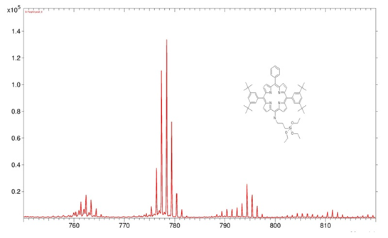
High mass range of a positive polarity secondary ion spectrum of a porphyrin derivative immobilized via silanol groups onto a silicon wafer (Bi3+). The multipletts reproduce the isotope distribution of this molecule and can be unambiguously assigned to the porphyrine headgroup, C54H58N5, etc.