
X-Ray Photoelectron Spectroscopy (XPS)
X-ray photoelectron spectroscopy (XPS) is the most widely used surface analysis technique to provide both quantitative atomic concentration and chemical state information of the detected elements. X-ray irradiation of surfaces results in the emission of photoelectrons whose energies are characteristic of the elements. The information depth is approximately 5–7 nm. Angle-resolved XPS offers non-destructive resolution of structures within the XPS sampling depth, e. g. layer ordering, composition and thickness can be determined. Moreover, XPS can be utilized for sputter depth profiling to characterize thin films and multi-layer systems by quantifying matrix-level elements as a function of depth. The gas cluster ion source additionally enables sputter depth profiling of organic materials while preserving the chemical information. The use of NAP XPS for non-vacuum-compatible samples is under development.
| Name | Phone | |
|---|---|---|
| Vanessa Trouillet | +49 721 608-22666 | vanessa trouillet ∂does-not-exist.kit edu |
| 1 additional person visible within KIT only. | ||
Details (XPS)
Features
K-Alpha & K-Alpha+ XPS Instruments
- Mono AlKα X-ray source, spot size 30–400 μm (spatial resolution)
- Energy resolution < 0.5 eV FWHM Ag 3d5/2
- Rapid snap map chemical imaging (K-Alpha+)
- Ion gun for sputter depth profiling, 100–3000 eV Ar+ ion energy (K-Alpha)
- Mono Atomic and Gas Cluster Ion Source (MAGCIS), Mono Ar+: 500 eV to 4 keV, Cluster Ar+: 2 keV to 8 keV, cluster size: 75 up to 2000 atoms (K-Alpha+)
- Charge neutralisation system
- 50 x 60 mm2 sample stage, sample height max. 15 mm
- Glove-box for atmosphere-contact free sample transfer: O2 < 1 ppm, H2O < 1 ppm (K-Alpha)
Escalab Xi+
- Mono AlKα X-ray source, spot size 200-900 µm, for elemental mapping down to 3 µm spatial resolution)
- MgKα/AlKα dual anode X-ray source
- Energy resolution < 0.85 eV FWHM Ag 3d5/2 (MgKα)
- Angle resolved XPS
- Mono Atomic and Gas Cluster Ion Source (MAGCIS),
Mono Ar+: 500 eV to 4 keV,
Cluster Ar+: 2 keV to 8 keV, cluster size: 75 up to 2000 atoms - Max. sample dimensions 20 x 30 x 10 mm2
- In-situ sample cooling and heating (-190–500 °C)
- UPS (He Source)
- REELS (1000 eV electron source)
- Glove-box (Argon) for inert gas sample transfer: O2 < 1 ppm, H2O < 1 ppm
Near Ambient Pressure XPS (under development)
- Mono AlKα X-ray source
- Up to a few mbars pressure in analysis chamber
- Gas loading possible
- Glove box for air/humidity sensitive samples
Limitations/constraints
- All elements are detectable except for H and He
- Sample has to be a solid at RT and stable under vacuum conditions, powders are possible. (Remark: NAP XPS brings some new possibilities.)
- Depending on the chemical composition samples might be sensitive to X-ray irradiation
Typical results
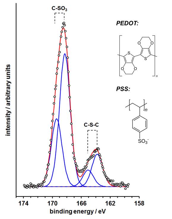
Fig. 1: Non-degraded S 2p XPS spectrum of a PEDOT:PSS thin layer after 1000 s Ar1000+ cluster ion sputtering using the K-Alpha+ MAGCIS source at 8 keV energy.
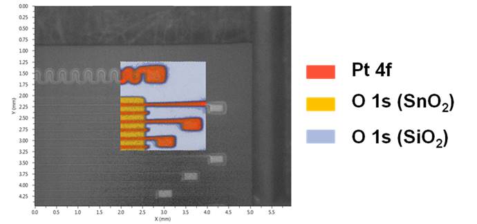
Fig. 2: Rapid snap map chemical image and respective overlaid video image of a gas sensor micro array (SiO2 chip substrate, SnO2 detector field subdivided by 50 µm Pt electrodes).
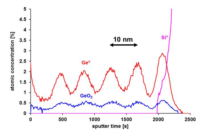
Fig. 3: Mono Ar+ ion sputter depth profile of a sputter deposited Ge-SiO2/SiO2 multilayer system.